
Introduction
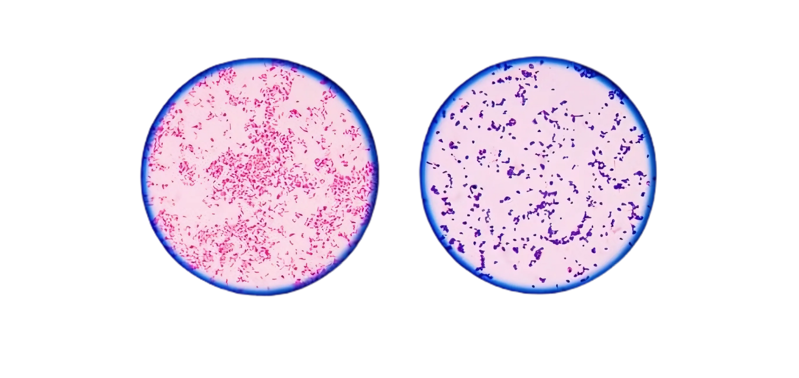
The gram stain test procedure highlights the differential staining characteristics of bacteria. Gram-positive bacteria retain the crystal violet dye after decolourization, appearing blue or purplish-blue under microscopy. In contrast, Gram-negative bacteria lose the crystal violet stain during decolourization and take up the counterstain (dilute carbol fuchsin), resulting in a red or pink appearance.
Table of Contents
Principle
pH Theory
Cytoplasm of gram-positive bacteria is more acidic, hence, can retain the basic dye (e.g. crystal violet) for longer time. Iodine serves as mordant, i.e. it combines with the primary stain to form a dye-iodine complex which gets retained inside the cell.
Cell wall theory
Gram-positive cell wall has a thick peptidoglycan layer (50–100 layers thick), with tight crlinkages
■ The peptidoglycan itself is not stained; instead, it seems to act as a permeability barpreventing loss of crystal violet. Large dye-iodine complexes are not able to penetrate ttightened peptidoglycan layerin a gram-positive bacteria. Gram-negative cell wall is more permeable thus allowing the outflow of crystal violet easily. Tis attributed to:
◆ The thin peptidoglycan layerin gram-negative cell wall which is not tightly cross linked
◆ Presence of lipopolysaccharide layer in the cell wall of gram-negative bacteria, whi
disrupted easily by the decolorizer; forming larger pores, that allow the dye-iodine compescape from the cytoplasm
Magnesium ribonucleate theory
It is present in the cell membrane of gram-positive bacteria but not in gram-negative bacteria and helps to retain the primary dye.
Check Out the Objectives of gram stain test procedure
- To determine and confirm the presence of bacteria and yeasts in clinical specimens using the Gram staining protocol.
- To establish the presence of potential pathogenic organisms, particularly in cases of suspected bacterial infections, where Gram’s stain plays a critical role in the initial evaluation (e.g., bacterial meningitis).
- To assess infection and contamination levels by examining Gram-stained smears for Pus cells, which indicate an active infection , squamous epithelial cells, which suggest mucosal or salivary contamination.
- To evaluate specimen quality by applying standardized criteria, such as rejecting sputum samples with >25 squamous epithelial cells per low-power field, as they indicate excessive saliva contamination and are unsuitable for bacterial culture.
- The Gram staining protocol serves as a rapid, cost-effective diagnostic tool to guide further microbiological testing and clinical decision-making.
sample Needed for gram stain test procedure
- Sputum, urine, and throat swabs
- Aspirate from various catheter
- swab from various locations
- CSF, pus, and other bodily fluids
- Tissue: Endometrium, bone marrow, biopsy material and surgical tissue and Semen
Gram Staining Modifications
- Jensen’s modification (useful for meningococci and gonococci)
- Brown and Brenn modification (used for Actinomycetes), etc.
Materials and Reagents
- Microslides
- Sterile normal saline
- Diamond marker
- Tissue paper
- Gram’s stain
- Immersion oil
Needed Equipment
- Laminar Air Flow Cabinet
- Microscope
Procedure
- Make a smear of the specimen on a clean labeled microslide and allow it to dry.
- Heat fix the smear to the slide by passing the slide 3 or 4 times through the flame of
Bunsen burner so that the smear does not wash off during the staining procedure. - Stain the slide by Gram’s staining procedure.
- Cover the smear with crystal violet and leave it for one minute.
- Wash the slide with water.
- Cover the slide with Gram’s iodine and leave it for one minute.
- Wash the slide with water.
- Decolorise quickly with acetone, rocking the slide gently.
- Wash with water immediately.
- Counterstain with dilute carbol fuchsin for 30 seconds.
- Wash with water and blot dry.
Interpretation of Results
- For sputum samples assess its quality by Bartlett’s score card given
- Grade and report vaginal smears as described by Nugent al given below.
- Mention the number of cells / oil immersion field for epithelial cells and pus
- Indicate the morphology and arrangement specifically, of the predominant bacteria either gram positive cocci or gram negative bacilli.
- Mention the coccobacillary or other forms if
Bartlett’s Grading System For Assessing Quality Of Sputum Samples
No. of Neutrophils per 10x low power field | Grade |
<10 | 0 |
10-25 | +1 |
>25 | +2 |
Presence of mucus | +1 |
No. of epithelial cells per 10x low power field | Grade |
10-25 | -1 |
>25 | -2 |
Average the number of epithelial neutrophils in about 20-30 separate 10x microscopic fields and then calculate the total. A final score 0 or less indicates lack of active inflammation or contamination with saliva. Repeat sample should be requested for.
Nugent’s Scoring Of Vaginal Swab For Diagnosis Of Bacterial Vaginosis
Interpretation of Neugent’s score | ||
Nugent’s score | And | Interpretation |
0-3 | No clue cells | Normal vaginal flora |
4-6 | No clue cells | Intermediate or Not consistent with Bacterial vaginosis |
4-6 | Clue cell present | Indicative of bacterial vaginosis |
≥ 7 | Clue cell present or absent | Indicative of bacterial vaginosis |
Quality Control Procedure
Check the Quality of stains with known ATCC stains of bacteria whenever new stains is used/prepared.